FONAR Upright™ MRI
|Clinical Images – FONAR Upright™ MRI
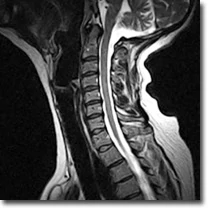
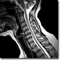
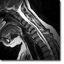
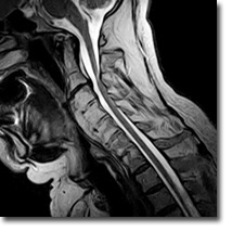
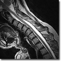
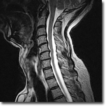
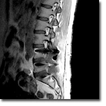
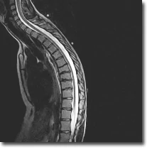
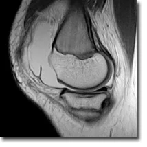
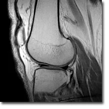
Sagittal sitting: hyperextension, neutral, flexion
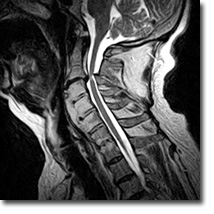
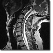
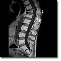
Sagittal sitting: neutral, flexion
Sagittal sitting: hyperextension, neutral, flexion
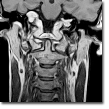

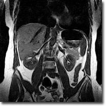
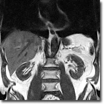
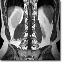
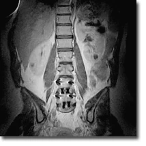
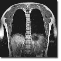
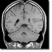
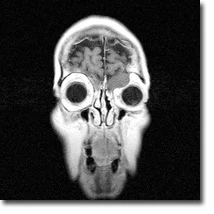
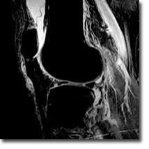
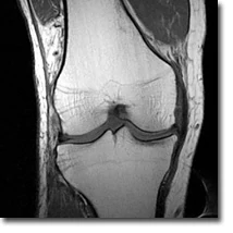
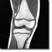
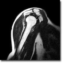
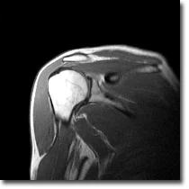
Coronal sitting
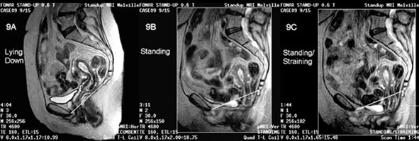
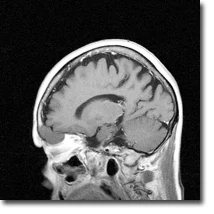
Standing: sagittal, transversal, coronal
Sitting: sagittal, transversal, coronal
Standing: sagittal, transversal, coronal
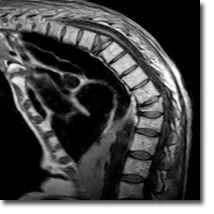
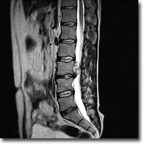
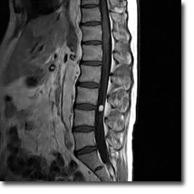
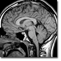
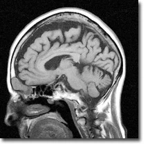
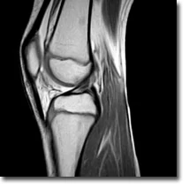
Sitting: sagittal
Sitting: sagittal, transversal,coronal
Sitting: sagittal, transversal,coronal
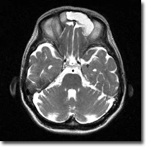

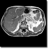
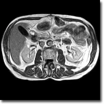
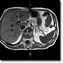
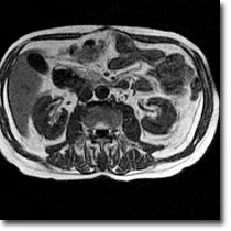
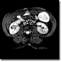
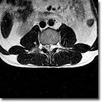
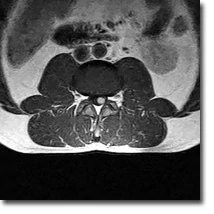
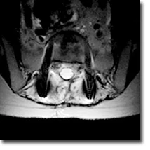
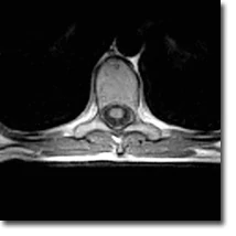
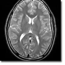
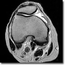
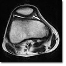
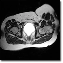
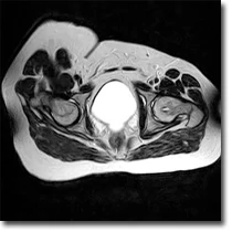
Sitting: transversal
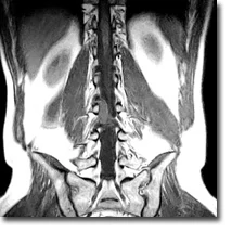
Sitting: sagittal, coronal
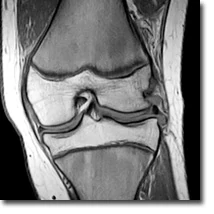
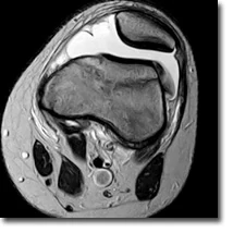

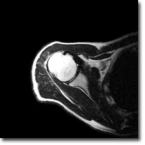
Imaging under weight bearing conditions: coronal, transversal, sagittal
Imaging under weight bearing conditions: sagittal, transversal, coronal
Imaging under weight bearing conditions: sagittal, transversal, coronal
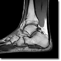
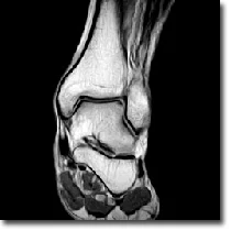
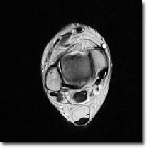
Imaging under weight bearing conditions: sagittal, coronal, transversal
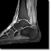
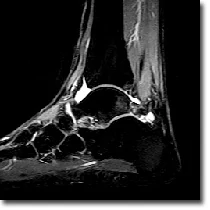
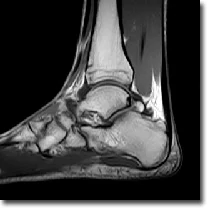
Imaging under weight bearing conditions: sagittal

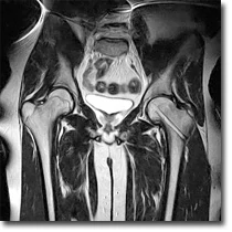
Standing on both legs: transversal, coronal
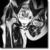
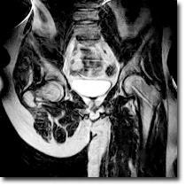
Standing on both legs: transversal, coronal
Standing